ab16325 APOA1 Antibody

Western blot analysis of extracts from various samples, using APOA1 Antibody. Lane 1: Rat brain lysates; Lane 2: Mouse brain lysates; Lane 3: Mouse muscle lysates; Lane 4: Mouse liver lysates;
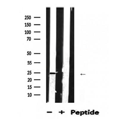
Western blot analysis of APOA1 expression in Mouse brain lysate.The lane on the right was treated with blocking peptide.
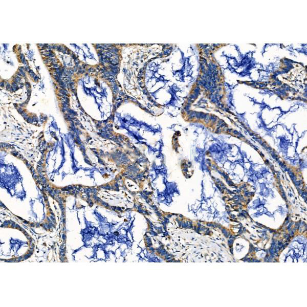
ab16325 at 1/100 staining Human colorectal cancer by IHC-P. The sample was formaldehyde fixed and a heat mediated antigen retrieval step in citrate buffer was performed. The sample was then blocked and incubated with the primary antibody at 4°C overnight. An HRP conjugated anti-Rabbit antibody was used as the secondary antibody.
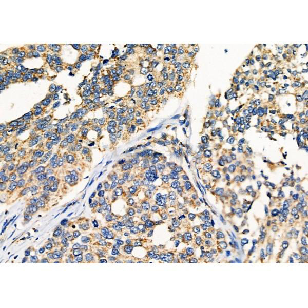
ab16325 at 1/100 staining Human ovarian cancer by IHC-P. The sample was formaldehyde fixed and a heat mediated antigen retrieval step in citrate buffer was performed. The sample was then blocked and incubated with the primary antibody at 4°C overnight. An HRP conjugated anti-Rabbit antibody was used as the secondary antibody.
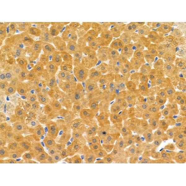
ab16325 at 1/100 staining Mouse liver tissue by IHC-P. The sample was formaldehyde fixed and a heat mediated antigen retrieval step in citrate buffer was performed. The sample was then blocked and incubated with the primary antibody at 4°C overnight. An HRP conjugated anti-Rabbit antibody was used as the secondary antibody.
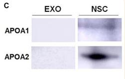
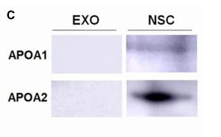
FIGURE 1 Characterization of exosomes (EXOs). (A) Purified exosomes were observed under transmission electron microscopy (TEM) using negative staining. (B) The levels of positive exosomal markers TSG101, CD9, and Flotillin-1 in protein lysates of neural stem cells (NSC)-derived exosome pellets were determined by western blotting. (C) The levels of negative exosomal markers APOA1 and APOA2 in protein lysates of NSC-derived exosome pellets and NSCs were determined by western blotting. (D,E) Particle-size distribution of exosomes was determined by NanoSight analysis (NTA) in panel (D) and NanoFCM in panel (E) technologies. Scale bar 200 nm in panel (A).
FIGURE 1 | Characterization of exosomes (EXOs). (C) The levels of negative exosomal markers APOA1 and APOA2 in protein lysates of NSC-derived exosome pellets and NSCs were determined by western blotting.
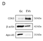
Fig. 3. Characterization of the extracellular vesicles (EVs) isolated from porcine follicular fluid. EVs size distribution (A), concentration (B) and zeta potential (C) were assessed by nanoparticle tracking analysis. Data is shown as the mean ± standard deviation (SD) (n = 6). Expression of CD63, β-actin and apolipoprotein A1 (Apo-A1) proteins in EVs samples and granulosa cell lysates (Gc) was demonstrated by Western blot (D). Immunofluorescence was used to confirm CD63 localization in porcine ovarian follicle (E). Positive signal (red) is marked by white arrows. Gc - granulosa cells; Ti - theca interna cells; Te -theca externa cells. (For interpretation of the references to colour in this figure legend, the reader is referred to the Web version of this article.)

品牌

产品货号

ab16325

来源种属

Rabbit

抗体克隆

Polyclonal

来源亚型

IgG

实验方法

WB,IHC

实验种属

Human,Mouse,Rat,Rabbit,Pig,Dog,Chicken,Bovine,Horse,Sheep

偶联标记

Unconjugated

目的蛋白

APOA1

产品规格

50μl,100μl,200μl

产品报价

¥1500/¥2750/¥3600

实验应用


Western blotting

Recommended dilution: 1:500-1:2000


Immunohistochemistry

Recommended dilution: 1:50-1:200


最佳稀释倍数与浓度应由实验研究人员确认

产品说明


产品背景

Participates in the reverse transport of cholesterol from tissues to the liver for excretion by promoting cholesterol efflux from tissues and by acting as a cofactor for the lecithin cholesterol acyltransferase (LCAT). As part of the SPAP complex, activates spermatozoa motility.

Description
Rabbit polyclonal antibody to APOA1

Applications 
WB, IHC.

Immunogen 
APOA1 Antibody detects endogenous levels of total APOA1.

Reactivity 
Human, Mouse, Rat.
可预测:Pig(100%), Bovine(%), Horse(%), Sheep(%), Rabbit(%), Dog(%)

Molecular weight
31kDa; 31kD(Calculated).

Host species 
Rabbit

Ig class 
Immunogen-specific rabbit IgG

Purification 
Antigen affinity purification

Full name 
APOA1

Synonyms 
Apo-AI; ApoA I; ApoA-I; APOA1; APOA1_HUMAN; Apolipoprotein A-I(1-242); Apolipoprotein A1; Apolipoprotein AI; Brp14; Ltw1; Lvtw1; Sep1; Sep2;

Storage
Rabbit IgG in phosphate buffered saline , pH 7.4, 150mM NaCl, 0.02% sodium azide and 50% glycerol. Store at -20 °C. Stable for 12 months from date of receipt.

Swissprot 
P02647

产品图片
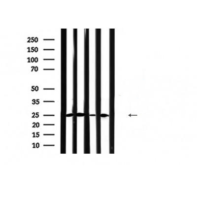
Western blot analysis of extracts from various samples, using APOA1 Antibody. Lane 1: Rat brain lysates; Lane 2: Mouse brain lysates; Lane 3: Mouse muscle lysates; Lane 4: Mouse liver lysates;

Western blot analysis of APOA1 expression in Mouse brain lysate.The lane on the right was treated with blocking peptide.

ab16325 at 1/100 staining Human colorectal cancer by IHC-P. The sample was formaldehyde fixed and a heat mediated antigen retrieval step in citrate buffer was performed. The sample was then blocked and incubated with the primary antibody at 4°C overnight. An HRP conjugated anti-Rabbit antibody was used as the secondary antibody.

ab16325 at 1/100 staining Human ovarian cancer by IHC-P. The sample was formaldehyde fixed and a heat mediated antigen retrieval step in citrate buffer was performed. The sample was then blocked and incubated with the primary antibody at 4°C overnight. An HRP conjugated anti-Rabbit antibody was used as the secondary antibody.

ab16325 at 1/100 staining Mouse liver tissue by IHC-P. The sample was formaldehyde fixed and a heat mediated antigen retrieval step in citrate buffer was performed. The sample was then blocked and incubated with the primary antibody at 4°C overnight. An HRP conjugated anti-Rabbit antibody was used as the secondary antibody.

FIGURE 1 Characterization of exosomes (EXOs). (A) Purified exosomes were observed under transmission electron microscopy (TEM) using negative staining. (B) The levels of positive exosomal markers TSG101, CD9, and Flotillin-1 in protein lysates of neural stem cells (NSC)-derived exosome pellets were determined by western blotting. (C) The levels of negative exosomal markers APOA1 and APOA2 in protein lysates of NSC-derived exosome pellets and NSCs were determined by western blotting. (D,E) Particle-size distribution of exosomes was determined by NanoSight analysis (NTA) in panel (D) and NanoFCM in panel (E) technologies. Scale bar 200 nm in panel (A).

FIGURE 1 | Characterization of exosomes (EXOs). (C) The levels of negative exosomal markers APOA1 and APOA2 in protein lysates of NSC-derived exosome pellets and NSCs were determined by western blotting.

Fig. 3. Characterization of the extracellular vesicles (EVs) isolated from porcine follicular fluid. EVs size distribution (A), concentration (B) and zeta potential (C) were assessed by nanoparticle tracking analysis. Data is shown as the mean ± standard deviation (SD) (n = 6). Expression of CD63, β-actin and apolipoprotein A1 (Apo-A1) proteins in EVs samples and granulosa cell lysates (Gc) was demonstrated by Western blot (D). Immunofluorescence was used to confirm CD63 localization in porcine ovarian follicle (E). Positive signal (red) is marked by white arrows. Gc - granulosa cells; Ti - theca interna cells; Te -theca externa cells. (For interpretation of the references to colour in this figure legend, the reader is referred to the Web version of this article.)

联系经销商
北京晶莱华科生物技术有限公司
北京总部电话:4008-766-085
邮箱:beijing@genlay.cn
地址:北京经济技术开发区科创十三街12号院德为科技园1号楼5层
上海分公司电话:4008-766-085
邮箱:shanghai@genlay.cn
地址:上海市青浦区普仓路648弄71号
长沙分公司电话:4008-766-085
邮箱:changsha@genlay.cn
地址:长沙市岳麓区麓谷大道627号新长海中心A1栋1203室
http://www.genlay.cn
http://www.genecro.cn
genetex@neobioscience.com