ab13159 Apelin Antibody
品牌 |
|
|---|---|
产品货号 |
ab13159 |
来源种属 |
Rabbit |
抗体克隆 |
Polyclonal |
来源亚型 |
IgG |
实验方法 |
WB,IHC,IF,ICC |
实验种属 |
Human,Mouse,Rat,Rabbit,Pig,Dog,Chicken,Bovine,Horse,Sheep |
偶联标记 |
Unconjugated |
目的蛋白 |
Apelin |
产品规格 |
50μl,100μl,200μl |
产品报价 |
¥1800/¥2750/¥3600 |
实验应用
Western blotting
Recommended dilution: 1:500-1:2000
Immunofluorescence
Recommended dilution: 1:100-1:500
immunocytochemistry
Recommended dilution: 1:100-1:500
Immunohistochemistry
最佳稀释倍数与浓度应由实验研究人员确认
产品说明
产品背景
Endogenous ligand for the apelin receptor (APLNR). Drives internalization of the apelin receptor (By similarity). Apelin-36 dissociates more hardly than (pyroglu)apelin-13 from APLNR (By similarity). Hormone involved in the regulation of cardiac precursor cell movements during gastrulation and heart morphogenesis (By similarity). Has an inhibitory effect on cytokine production in response to T-cell receptor/CD3 cross-linking; the oral intake of apelin in the colostrum and the milk might therefore modulate immune responses in neonates (By similarity). Plays a role in early coronary blood vessels formation (By similarity). Mediates myocardial contractility in an ERK1/2-dependent manner (By similarity). May also have a role in the central control of body fluid homeostasis by influencing vasopressin release and drinking behavior (By similarity).
(Microbial infection) Endogenous ligand for the apelin receptor (APLNR), an alternative coreceptor with CD4 for HIV-1 infection. Inhibits HIV-1 entry in cells coexpressing CD4 and APLNR. Apelin-36 has a greater inhibitory activity on HIV infection than other synthetic apelin derivatives.
Description
Rabbit polyclonal antibody to Apelin
Applications
WB, IF, ICC, IHC.
Immunogen
Apelin Antibody detects endogenous levels of total Apelin.
Reactivity
Human, Mouse, Rat.
可预测:Pig(100%), Bovine(%), Sheep(%), Rabbit(%)
Molecular weight
8kDa; 9kD(Calculated).
Host species
Rabbit
Ig class
Immunogen-specific rabbit IgG
Purification
Antigen affinity purification
Full name
Apelin
Synonyms
AGTRL1 ligand; APEL; APEL_HUMAN; Apelin-13; APJ endogenous ligand; Apln; XNPEP2;
Storage
Rabbit IgG in phosphate buffered saline , pH 7.4, 150mM NaCl, 0.02% sodium azide and 50% glycerol. Store at -20 °C. Stable for 12 months from date of receipt.
Swissprot
Q9ULZ1
产品图片

|
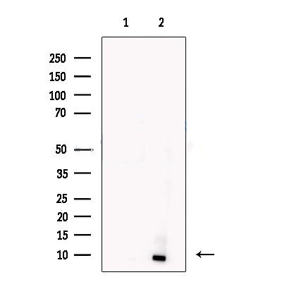
Western blot analysis of extracts from Hela cells, using Apelin Antibody. The lane on the left was treated with blocking peptide. |


![Figure 5. Immunohistochemical analysis of CD34, vascular endothelial growth factor (VEGF), and angiogenesis-related differentially expressed genes in the experimental (alveolar echinococcosis [AE]) and control (AE control group [AEC]) group samples. (A) CD34 and VEGF staining in AE samples showed different degrees of vascularization. Increased expression of SPP1, RSPO3, APLN, TWIST1, ADAM12, and FOXC2 was observed in the AE samples, whereas low or no expression of these genes was observed in the AEC samples. (B) The H-scores of CD34, VEGF, SPP1, RSPO3, APLN, TWIST1, ADAM12, and FOXC2 were significantly higher in the AE samples than in the AEC samples (values represent the mean ± standard error of the mean: ∗∗∗P < .001, ∗∗∗∗P < .0001). Figure 5. Immunohistochemical analysis of CD34, vascular endothelial growth factor (VEGF), and angiogenesis-related differentially expressed genes in the experimental (alveolar echinococcosis [AE]) and control (AE control group [AEC]) group samples. (A) CD34 and VEGF staining in AE samples showed different degrees of vascularization. Increased expression of SPP1, RSPO3, APLN, TWIST1, ADAM12, and FOXC2 was observed in the AE samples, whereas low or no expression of these genes was observed in the AEC samples. (B) The H-scores of CD34, VEGF, SPP1, RSPO3, APLN, TWIST1, ADAM12, and FOXC2 were significantly higher in the AE samples than in the AEC samples (values represent the mean ± standard error of the mean: ∗∗∗P < .001, ∗∗∗∗P < .0001).](/uploads/allimg/250808/1-1K461E64-3001.png)

