ab16420 AMPK alpha Antibody

Western blot analysis of extracts from PANC1, using AMPK alpha Antibody. The lane on the left was treated with blocking peptide.
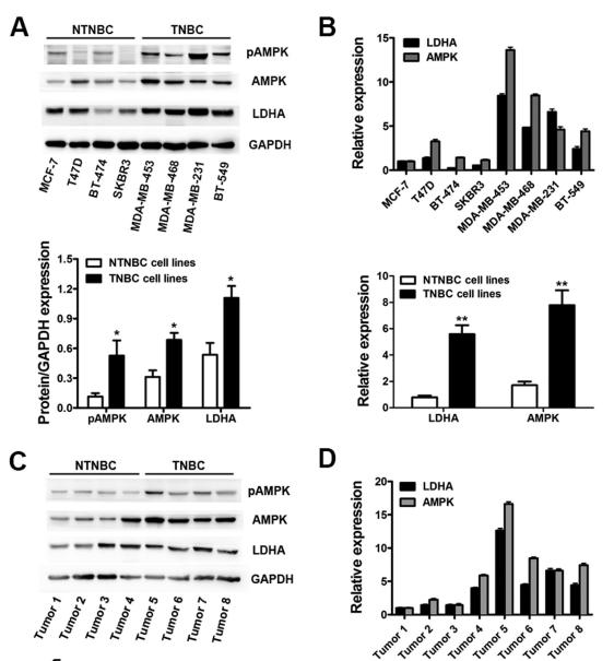
Fig. 1. LDHA and AMPK were up-regulated synchronously in breast cancer. A. Expression levels of LDHA, AMPK and pAMPK were assessed by Western blot (above) and gray image scanning (below) in eight different breast cancer cell lines, including four NTNBC cell lines and four TNBC cell lines. GAPDH was used as a loading control. B. Expression levels of LDHA and AMPK were determined by qRT-PCR (above). The differences between TNBC and NTNBC cell lines were analyzed (below). GAPDH was used as an internal control. C. Expression levels of LDHA, AMPK and pAMPK were assessed by Western blot (above) and gray image scanning (below) in eight different breast cancer tissues, including four NTNBC tissues and four TNBC tissues. GAPDH was used as a loading control. D. Expression levels of LDHA and AMPK were determined by qRT-PCR (above). The diffe
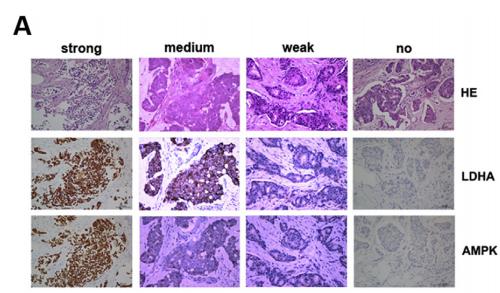
Fig. 2. The expression of LDHA and AMPK showed a positive correlation in breast cancer. A. The expression of LDHA and AMPK were detected by IHC using breast cancer TMAs of 112 patients. Representative IHC images of four staining degrees (no-weak-medium-strong) of LDHA and AMPK expression under a microscope were showed (400). B. The correlation between LDHA and AMPK expression scores of 112 breast cancer patients was analyzed and a positive correlation between them was showed.
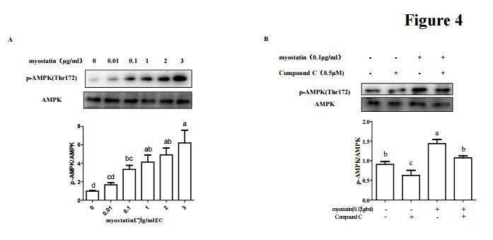
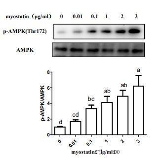
Fig. 4. Myostatin regulated translation elongation through AMP. C2C12 myotubes were treated with various concentration recombinant myostatin (0, 0.01,0.1, 1, 2, 3 µg/ml) for 48 h andthen lysed and cellular extracts were analyzed by Western blot with anti-AMPK(A).
Fig. 4. |Myostatin regulated translation elongation through AMPK.C2C12 myotubes were treated with various concentration recombinant myostatin (0, 0.01,429 0.1, 1, 2, 3 µg/ml) for 48 h andthen lysed and cellular extracts were analyzed by Western blot with 430 anti-AMPK(A).
Fig. 5| AA treatment activated the AMPK and inhibited PI3K/AKT signaling pathway. The cells were treated with or without AA (5 μM) for 3 days. aRepresentative western blot of p-AMPK, AMPK, p-PI3K, PI3K, p-AKT, and AKT. GAPDH was served as a loading control.
Fig. 5 AA treatment activated the AMPK and inhibited PI3K/AKT signaling pathway. The cells were treated with or without AA (5 μM) for 3 days. a Representative western blot of p-AMPK, AMPK, p-PI3K, PI3K, p-AKT, and AKT. GAPDH was served as a loading control. b Relative protein expression of p-AMPK/AMPK, p-PI3K/PI3K, and p-AKT/AKT. Unpaired t test, *P < 0.05, ***P < 0.001. Each experiment was repeated three times (p-AMPK, phosphorylated AMP-activated protein kinase; AMPK, AMP-activated protein kinase; p-PI3K, phosphorylated phosphoinositide-3 kinase; PI3K, phosphoinositide-3 kinase; p-AKT, phosphorylated protein kinase B; AKT, protein kinase B)
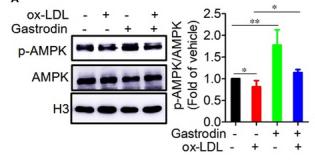
FIGURE 6 AMPK is a critical upstream regulator of FoxO1 and TFEB. A and B, Gastrodin activated AMPK in the foam cells. A, Representative blots of AMPK and p‐AMPK in macrophages. B, Immunofluorescence analysis of p‐AMPK in macrophages. C and D, The inhibition of AMPK activity decreased the phosphorylation of FoxO1 and nuclear translocation of TFEB. Macrophages were treated with CC (10μM) for 1 h. The phosphorylation level of FoxO1 was analysed by Western blotting C, and nuclear translocation of TFEB was determined by immunofluorescence D. *P < .05; **P < .01. Results are presented as mean ± SD of three independent experiments. The value represents fold of vehicle. CC, Dorsomorphin dihydrochloride

品牌

产品货号

ab16420

来源种属

Rabbit

抗体克隆

Polyclonal

来源亚型

IgG

实验方法

WB,IHC,IF,ICC

实验种属

Human,Mouse,Rat,Rabbit,Pig,Dog,Chicken,Bovine,Horse,Sheep

偶联标记

Unconjugated

目的蛋白

AMPK alpha

产品规格

50μl,100μl,200μl

产品报价

¥1500/¥2750/¥3600

实验应用


Western blotting

Recommended dilution: 1:500-1:2000


Immunofluorescence

Recommended dilution: 1:100


immunocytochemistry

Recommended dilution: 1:100


Immunohistochemistry

Recommended dilution: 1:50-1:200


最佳稀释倍数与浓度应由实验研究人员确认

产品说明


产品背景

Catalytic subunit of AMP-activated protein kinase (AMPK), an energy sensor protein kinase that plays a key role in regulating cellular energy metabolism. In response to reduction of intracellular ATP levels, AMPK activates energy-producing pathways and inhibits energy-consuming processes: inhibits protein, carbohydrate and lipid biosynthesis, as well as cell growth and proliferation. AMPK acts via direct phosphorylation of metabolic enzymes, and by longer-term effects via phosphorylation of transcription regulators. Also acts as a regulator of cellular polarity by remodeling the actin cytoskeleton; probably by indirectly activating myosin. Regulates lipid synthesis by phosphorylating and inactivating lipid metabolic enzymes such as ACACA, ACACB, GYS1, HMGCR and LIPE; regulates fatty acid and cholesterol synthesis by phosphorylating acetyl-CoA carboxylase (ACACA and ACACB) and hormone-sensitive lipase (LIPE) enzymes, respectively. Regulates insulin-signaling and glycolysis by phosphorylating IRS1, PFKFB2 and PFKFB3. AMPK stimulates glucose uptake in muscle by increasing the translocation of the glucose transporter SLC2A4/GLUT4 to the plasma membrane, possibly by mediating phosphorylation of TBC1D4/AS160. Regulates transcription and chromatin structure by phosphorylating transcription regulators involved in energy metabolism such as CRTC2/TORC2, FOXO3, histone H2B, HDAC5, MEF2C, MLXIPL/ChREBP, EP300, HNF4A, p53/TP53, SREBF1, SREBF2 and PPARGC1A. Acts as a key regulator of glucose homeostasis in liver by phosphorylating CRTC2/TORC2, leading to CRTC2/TORC2 sequestration in the cytoplasm. In response to stress, phosphorylates 'Ser-36' of histone H2B (H2BS36ph), leading to promote transcription. Acts as a key regulator of cell growth and proliferation by phosphorylating TSC2, RPTOR and ATG1/ULK1: in response to nutrient limitation, negatively regulates the mTORC1 complex by phosphorylating RPTOR component of the mTORC1 complex and by phosphorylating and activating TSC2. In response to nutrient limitation, promotes autophagy by phosphorylating and activating ATG1/ULK1. In that process also activates WDR45. In response to nutrient limitation, phosphorylates transcription factor FOXO3 promoting FOXO3 mitochondrial import (By similarity). AMPK also acts as a regulator of circadian rhythm by mediating phosphorylation of CRY1, leading to destabilize it. May regulate the Wnt signaling pathway by phosphorylating CTNNB1, leading to stabilize it. Also has tau-protein kinase activity: in response to amyloid beta A4 protein (APP) exposure, activated by CAMKK2, leading to phosphorylation of MAPT/TAU; however the relevance of such data remains unclear in vivo. Also phosphorylates CFTR, EEF2K, KLC1, NOS3 and SLC12A1.

Description
Rabbit polyclonal antibody to AMPK alpha

Applications 
WB, IF, ICC, IHC.

Immunogen 
AMPK alpha Antibody detects endogenous levels of total AMPK alpha.

Reactivity 
Human, Mouse, Rat.
可预测:Zebrafish(88%), Bovine(%), Sheep(%), Rabbit(%), Dog(%), Chicken(%)

Molecular weight
63kDa; 64kD,62kD(Calculated).

Host species 
Rabbit

Ig class 
Immunogen-specific rabbit IgG

Purification 
Antigen affinity purification

Full name 
AMPK alpha

Synonyms 
5 AMP activated protein kinase alpha 1catalytic subunit; 5 AMP activated protein kinase catalytic alpha 1 chain; 5' AMP activated protein kinase catalytic subunit alpha 1; 5'-AMP-activated protein kinase catalytic subunit alpha-1; AAPK1; AAPK1_HUMAN; ACACA kinase; acetyl CoA carboxylase kinase; AI194361; AI450832; AL024255; AMP -activate kinase alpha 1 subunit; AMP-activated protein kinase, catalytic, alpha -1; AMPK 1; AMPK alpha 1; AMPK alpha 1 chain; AMPK; AMPK subunit alpha-1; AMPK1; AMPKa1; AMPKalpha1; C130083N04Rik; cb116; EC 2.7.11.1; HMG CoA reductase kinase; HMGCR kinase; hormone sensitive lipase kinase; Hydroxymethylglutaryl CoA reductase kinase; im:7154392; kinase AMPK alpha1; MGC33776; MGC57364; OTTHUMP00000161795; OTTHUMP00000161796; PRKAA 1; PRKAA1; Protein kinase AMP activated alpha 1 catalytic subunit; SNF1-like protein AMPK; SNF1A; Tau protein kinase PRKAA1; wu:fa94c10; 5'-AMP-activated protein kinase catalytic subunit alpha-2; AAPK2_HUMAN; ACACA kinase; Acetyl-CoA carboxylase kinase; AMPK alpha 2 chain; AMPK subunit alpha-2; AMPK2; AMPKa2; AMPKalpha2; HMGCR kinase; Hydroxymethylglutaryl-CoA reductase kinase; PRKAA; PRKAA2; Protein kinase AMP activated alpha 2 catalytic subunit; Protein kinase AMP activated catalytic subunit alpha 2;

Storage
Rabbit IgG in phosphate buffered saline , pH 7.4, 150mM NaCl, 0.02% sodium azide and 50% glycerol. Store at -20 °C. Stable for 12 months from date of receipt.

Swissprot 
Q13131 | P54646

产品图片
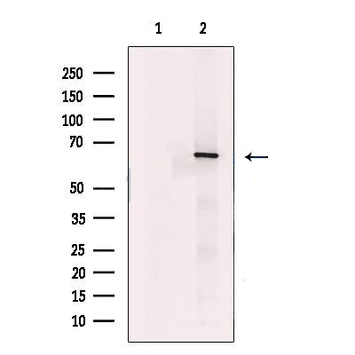
Western blot analysis of extracts from PANC1, using AMPK alpha Antibody. The lane on the left was treated with blocking peptide.

Fig. 1. LDHA and AMPK were up-regulated synchronously in breast cancer. A. Expression levels of LDHA, AMPK and pAMPK were assessed by Western blot (above) and gray image scanning (below) in eight different breast cancer cell lines, including four NTNBC cell lines and four TNBC cell lines. GAPDH was used as a loading control. B. Expression levels of LDHA and AMPK were determined by qRT-PCR (above). The differences between TNBC and NTNBC cell lines were analyzed (below). GAPDH was used as an internal control. C. Expression levels of LDHA, AMPK and pAMPK were assessed by Western blot (above) and gray image scanning (below) in eight different breast cancer tissues, including four NTNBC tissues and four TNBC tissues. GAPDH was used as a loading control. D. Expression levels of LDHA and AMPK were determined by qRT-PCR (above). The diffe

Fig. 2. The expression of LDHA and AMPK showed a positive correlation in breast cancer. A. The expression of LDHA and AMPK were detected by IHC using breast cancer TMAs of 112 patients. Representative IHC images of four staining degrees (no-weak-medium-strong) of LDHA and AMPK expression under a microscope were showed (400). B. The correlation between LDHA and AMPK expression scores of 112 breast cancer patients was analyzed and a positive correlation between them was showed.

Fig. 4. Myostatin regulated translation elongation through AMP. C2C12 myotubes were treated with various concentration recombinant myostatin (0, 0.01,0.1, 1, 2, 3 µg/ml) for 48 h andthen lysed and cellular extracts were analyzed by Western blot with anti-AMPK(A).

Fig. 4. |Myostatin regulated translation elongation through AMPK.C2C12 myotubes were treated with various concentration recombinant myostatin (0, 0.01,429 0.1, 1, 2, 3 µg/ml) for 48 h andthen lysed and cellular extracts were analyzed by Western blot with 430 anti-AMPK(A).

Fig. 5| AA treatment activated the AMPK and inhibited PI3K/AKT signaling pathway. The cells were treated with or without AA (5 μM) for 3 days. aRepresentative western blot of p-AMPK, AMPK, p-PI3K, PI3K, p-AKT, and AKT. GAPDH was served as a loading control.

Fig. 5 AA treatment activated the AMPK and inhibited PI3K/AKT signaling pathway. The cells were treated with or without AA (5 μM) for 3 days. a Representative western blot of p-AMPK, AMPK, p-PI3K, PI3K, p-AKT, and AKT. GAPDH was served as a loading control. b Relative protein expression of p-AMPK/AMPK, p-PI3K/PI3K, and p-AKT/AKT. Unpaired t test, *P < 0.05, ***P < 0.001. Each experiment was repeated three times (p-AMPK, phosphorylated AMP-activated protein kinase; AMPK, AMP-activated protein kinase; p-PI3K, phosphorylated phosphoinositide-3 kinase; PI3K, phosphoinositide-3 kinase; p-AKT, phosphorylated protein kinase B; AKT, protein kinase B)

FIGURE 6 AMPK is a critical upstream regulator of FoxO1 and TFEB. A and B, Gastrodin activated AMPK in the foam cells. A, Representative blots of AMPK and p‐AMPK in macrophages. B, Immunofluorescence analysis of p‐AMPK in macrophages. C and D, The inhibition of AMPK activity decreased the phosphorylation of FoxO1 and nuclear translocation of TFEB. Macrophages were treated with CC (10μM) for 1 h. The phosphorylation level of FoxO1 was analysed by Western blotting C, and nuclear translocation of TFEB was determined by immunofluorescence D. *P < .05; **P < .01. Results are presented as mean ± SD of three independent experiments. The value represents fold of vehicle. CC, Dorsomorphin dihydrochloride

联系经销商
北京晶莱华科生物技术有限公司
北京总部电话:4008-766-085
邮箱:beijing@genlay.cn
地址:北京经济技术开发区科创十三街12号院德为科技园1号楼5层
上海分公司电话:4008-766-085
邮箱:shanghai@genlay.cn
地址:上海市青浦区普仓路648弄71号
长沙分公司电话:4008-766-085
邮箱:changsha@genlay.cn
地址:长沙市岳麓区麓谷大道627号新长海中心A1栋1203室
http://www.genlay.cn
http://www.genecro.cn
genetex@neobioscience.com