ab16286 Alkaline Phosphatase Antibody

Western blot analysis of ALPL expression in Mouse brain lysates
Fig. 5 |ADSC-Exos improved the healing of tendon injury. a, h The H&E staining of tendon injury at week 2 and week 4. b–f, i–m The expression of TNMD, collagen I, SCXA, ALP, and Runx2 were detected by immunohistochemistry assay at week 2 (n = 6) and week 4 (n = 6).
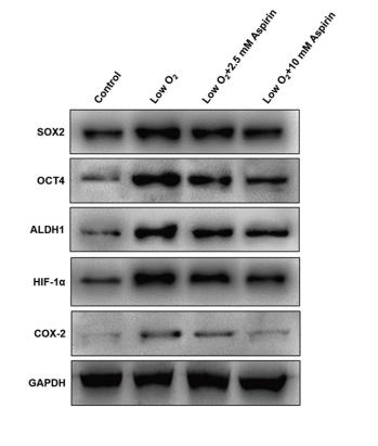
Fig. 2.| Aspirin inhibits the hypoxia-enhanced stemness of A549 cells. (A–C) mRNA levels of SOX-2, OCT4, and ALDH1 were determined by qRT–PCR using specific primers, with GAPDH serving as the internal control. (D, E) mRNA levels of HIF-1α and COX-2 were determined by qRT–PCR using specific primers, with GAPDH serving as the internal control. (F) The concentration of PGE2 secreted by A549 cells was determined by ELISA. Each bar indicates the mean ± SD of n = 3 experiments. * indicates P < 0.05, ** indicates P < 0.01. (G) SOX-2, OCT4, ALDH1, HIF-1α, and COX-2 protein levels were determined by Western blot, with GAPDH serving as the loading control.
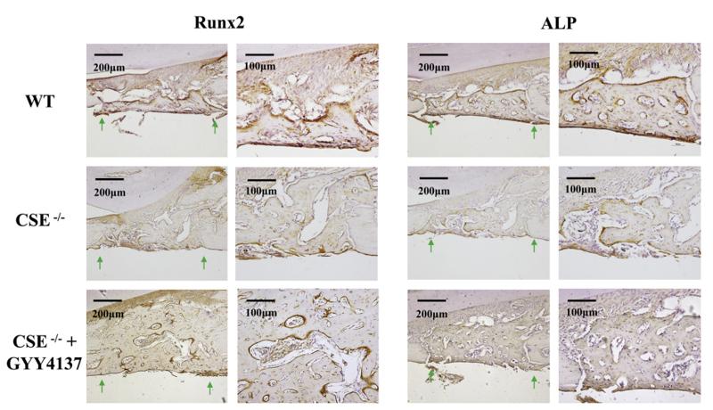
Fig. 4. Immunohistochemistry stainings of RUNX2 and ALP in the mandibular defect area of WT, CSE−/−and CSE−/− + GYY4137 mice 2 weeks after surgery. WT and CSE−/− + GYY4137 mice demonstrated increased expression levels of RUNX2 and ALP in the defect area 2 weeks after surgery. Green arrows represent the margin of the mandibular defect area. WT: wild type; CSE-/-: CSE knock out mice; Runx2: runt-related transcription factor 2; ALP: alkaline phosphatase. (For interpretation of the references to colour in this figure legend, the reader is referred to the web version of this article).
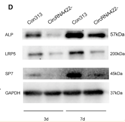
Figure 3 circRNA422 interfering inhibited osteogenic differentiation of BMSCs (A) ALP staining to evaluate the effect of circRNA422− on the ALP activity of BMSCs on osteogenic days 3 and 7. (B) Protein levels of OCN to evaluate the effect of circRNA422− on mineralization of BMSCs on osteogenic days 14 and 21. (C) Quantitative real-time PCR analysis showed that the osteogenic mRNA expressions levels of SP7, LRP5, ALP, OCN, BSP, and OPN were downregulated by circRNA422− compared with Con313 on osteogenic days 3 and 7 (n = 3, ∗p < 0.05). (D) WB analysis showed that the osteogenic protein expressions levels of ALP, LRP5, and SP7 were reduced by circRNA422− compared with Con313 on osteogenic days 3 and 7 (n = 3, ∗p < 0.05).
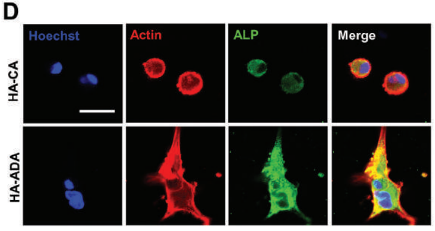
Figure 7. The dynamic HA-ADA hydrogel enhances the osteogenic differentiation. D) Representative images and E) quantified intensity (n = 20) of ALP (green) immunostaining of the hMSCs in the hydrogels.
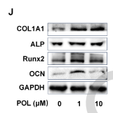
Figure 1. POL stimulated BMSCs proliferation and osteogenic differentiation. (A) POL chemical structure; (B) BMSCs surface markers were detected by flow cytometry; (C) The result of CCK-8 assay (n = 5); (D-E) The result of CFU assay (n = 5); (F) Images of ALP staining (scale bar = 250 μm); (G) Quantification of ALP staining (n = 5); (H) Images of ARS staining (scale bar = 250 μm); (I) Quantification of ARS staining (n = 5); (J-K) The protein expression levels of OCN, RUNX2, ALP, and COL1A1 were evaluated by western blot (n = 3); (L-M) Immunofluorescence staining of OCN and ALP (scale bar = 200 μm). Data were presented as mean ± SEM. Compared with control group: * P < 0.05, **P < 0.01, ***P < 0.001. Compared with 1 μM group: # P < 0.05, ##P < 0.01, ###P < 0.001.

品牌

产品货号

来源种属

Rabbit

抗体克隆

Polyclonal

来源亚型

IgG

实验方法

WB,IHC,IF,ICC

实验种属

Human,Mouse,Rat,Rabbit,Pig,Dog,Chicken,Bovine,Horse,Sheep

偶联标记

Unconjugated

目的蛋白

Alkaline Phosphatase

产品规格

50μl,100μl,200μl

产品报价

¥1500/¥2750/¥3600

实验应用

Western blotting

Recommended dilution: 1:500-1:2000


Immunofluorescence

Recommended dilution: 1:100-1:500


immunocytochemistry

Recommended dilution: 1:100-1:500


Immunohistochemistry

Recommended dilution: 1:50-1:200


最佳稀释倍数与浓度应由实验研究人员确认

产品说明


产品背景

This isozyme plays a key role in skeletal mineralization by regulating levels of diphosphate (PPi).

Description
Rabbit polyclonal antibody to Alkaline Phosphatase

Applications 
WB, IF, ICC, IHC.

Immunogen 
ALPL Antibody detects endogenous levels of total ALPL.

Reactivity 
Human, Mouse, Rat.
可预测:Pig(88%), Bovine(%), Horse(%), Sheep(%), Rabbit(%), Dog(%)

Molecular weight
57kDa; 57kD(Calculated).

Host species 
Rabbit

Ig class 
Immunogen-specific rabbit IgG

Purification 
Antigen affinity purification

Full name 
Alkaline Phosphatase

Synonyms 
AKP2; Alkaline phosphatase liver/bone/kidney; Alkaline phosphatase liver/bone/kidney isozyme; Alkaline phosphatase tissue nonspecific isozyme; Alkaline phosphatase, tissue-nonspecific isozyme; Alkaline phosphomonoesterase; Alpl; AP TNAP; AP-TNAP; APTNAP; BAP; FLJ40094; FLJ93059; Glycerophosphatase; HOPS; Liver/bone/kidney type alkaline phosphatase; MGC161443; MGC167935; PHOA; PPBT_HUMAN; Tissue non specific alkaline phosphatase; Tissue nonspecific ALP; TNAP; TNSALP;

Storage
Rabbit IgG in phosphate buffered saline , pH 7.4, 150mM NaCl, 0.02% sodium azide and 50% glycerol. Store at -20 °C. Stable for 12 months from date of receipt.

Swissprot 
P05186

产品图片
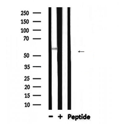
Western blot analysis of ALPL expression in Mouse brain lysates

Fig. 5 |ADSC-Exos improved the healing of tendon injury. a, h The H&E staining of tendon injury at week 2 and week 4. b–f, i–m The expression of TNMD, collagen I, SCXA, ALP, and Runx2 were detected by immunohistochemistry assay at week 2 (n = 6) and week 4 (n = 6).

Fig. 2.| Aspirin inhibits the hypoxia-enhanced stemness of A549 cells. (A–C) mRNA levels of SOX-2, OCT4, and ALDH1 were determined by qRT–PCR using specific primers, with GAPDH serving as the internal control. (D, E) mRNA levels of HIF-1α and COX-2 were determined by qRT–PCR using specific primers, with GAPDH serving as the internal control. (F) The concentration of PGE2 secreted by A549 cells was determined by ELISA. Each bar indicates the mean ± SD of n = 3 experiments. * indicates P < 0.05, ** indicates P < 0.01. (G) SOX-2, OCT4, ALDH1, HIF-1α, and COX-2 protein levels were determined by Western blot, with GAPDH serving as the loading control.

Fig. 4. Immunohistochemistry stainings of RUNX2 and ALP in the mandibular defect area of WT, CSE−/−and CSE−/− + GYY4137 mice 2 weeks after surgery. WT and CSE−/− + GYY4137 mice demonstrated increased expression levels of RUNX2 and ALP in the defect area 2 weeks after surgery. Green arrows represent the margin of the mandibular defect area. WT: wild type; CSE-/-: CSE knock out mice; Runx2: runt-related transcription factor 2; ALP: alkaline phosphatase. (For interpretation of the references to colour in this figure legend, the reader is referred to the web version of this article).

Figure 3 circRNA422 interfering inhibited osteogenic differentiation of BMSCs (A) ALP staining to evaluate the effect of circRNA422− on the ALP activity of BMSCs on osteogenic days 3 and 7. (B) Protein levels of OCN to evaluate the effect of circRNA422− on mineralization of BMSCs on osteogenic days 14 and 21. (C) Quantitative real-time PCR analysis showed that the osteogenic mRNA expressions levels of SP7, LRP5, ALP, OCN, BSP, and OPN were downregulated by circRNA422− compared with Con313 on osteogenic days 3 and 7 (n = 3, ∗p < 0.05). (D) WB analysis showed that the osteogenic protein expressions levels of ALP, LRP5, and SP7 were reduced by circRNA422− compared with Con313 on osteogenic days 3 and 7 (n = 3, ∗p < 0.05).

Figure 7. The dynamic HA-ADA hydrogel enhances the osteogenic differentiation. D) Representative images and E) quantified intensity (n = 20) of ALP (green) immunostaining of the hMSCs in the hydrogels.

Figure 1. POL stimulated BMSCs proliferation and osteogenic differentiation. (A) POL chemical structure; (B) BMSCs surface markers were detected by flow cytometry; (C) The result of CCK-8 assay (n = 5); (D-E) The result of CFU assay (n = 5); (F) Images of ALP staining (scale bar = 250 μm); (G) Quantification of ALP staining (n = 5); (H) Images of ARS staining (scale bar = 250 μm); (I) Quantification of ARS staining (n = 5); (J-K) The protein expression levels of OCN, RUNX2, ALP, and COL1A1 were evaluated by western blot (n = 3); (L-M) Immunofluorescence staining of OCN and ALP (scale bar = 200 μm). Data were presented as mean ± SEM. Compared with control group: * P < 0.05, **P < 0.01, ***P < 0.001. Compared with 1 μM group: # P < 0.05, ##P < 0.01, ###P < 0.001.

联系经销商
北京晶莱华科生物技术有限公司
北京总部电话:4008-766-085
邮箱:beijing@genlay.cn
地址:北京经济技术开发区科创十三街12号院德为科技园1号楼5层
上海分公司电话:4008-766-085
邮箱:shanghai@genlay.cn
地址:上海市青浦区普仓路648弄71号
长沙分公司电话:4008-766-085
邮箱:changsha@genlay.cn
地址:长沙市岳麓区麓谷大道627号新长海中心A1栋1203室
http://www.genlay.cn
http://www.genecro.cn
genetex@neobioscience.com