ab17521 Aggrecan Antibody

Western blot analysis of extracts from 3T3, using Aggrecan Antibody. The lane on the left was treated with blocking peptide.
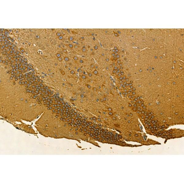
ab17521 at 1/100 staining Rat brain tissue by IHC-P. The sample was formaldehyde fixed and a heat mediated antigen retrieval step in citrate buffer was performed. The sample was then blocked and incubated with the primary antibody at 4°C overnight. An HRP conjugated anti-Rabbit antibody was used as the secondary antibody.
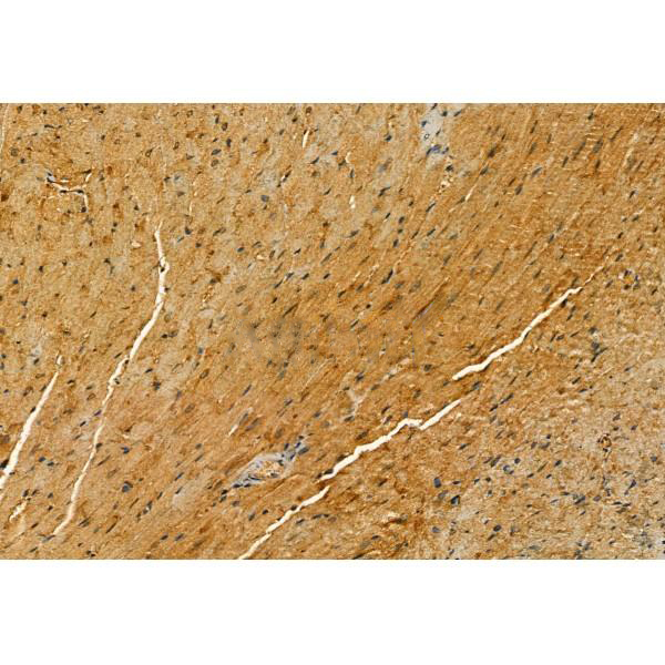
ab17521 at 1/100 staining Mouse heart tissue by IHC-P. The sample was formaldehyde fixed and a heat mediated antigen retrieval step in citrate buffer was performed. The sample was then blocked and incubated with the primary antibody at 4°C overnight. An HRP conjugated anti-Rabbit antibody was used as the secondary antibody.
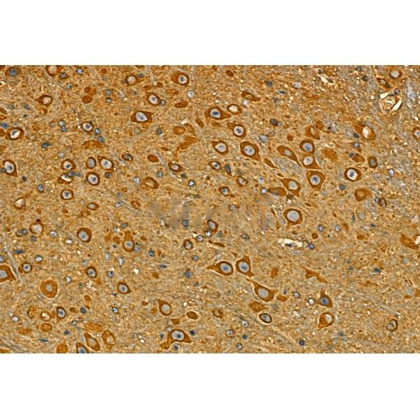
ab17521 at 1/100 staining Mouse brain tissue by IHC-P. The sample was formaldehyde fixed and a heat mediated antigen retrieval step in citrate buffer was performed. The sample was then blocked and incubated with the primary antibody at 4°C overnight. An HRP conjugated anti-Rabbit antibody was used as the secondary antibody.
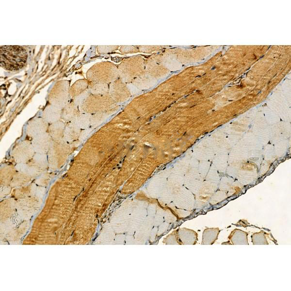
ab17521 at 1/100 staining Mouse skin tissue by IHC-P. The sample was formaldehyde fixed and a heat mediated antigen retrieval step in citrate buffer was performed. The sample was then blocked and incubated with the primary antibody at 4°C overnight. An HRP conjugated anti-Rabbit antibody was used as the secondary antibody.
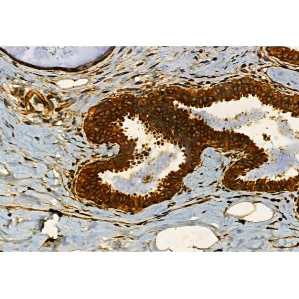
ab17521 at 1/100 staining Human normal tissues adjacent to mammary cancer by IHC-P. The sample was formaldehyde fixed and a heat mediated antigen retrieval step in citrate buffer was performed. The sample was then blocked and incubated with the primary antibody at 4°C overnight. An HRP conjugated anti-Rabbit antibody was used as the secondary antibody.
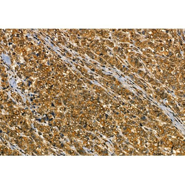
ab17521 at 1/100 staining Human mammary cancer by IHC-P. The sample was formaldehyde fixed and a heat mediated antigen retrieval step in citrate buffer was performed. The sample was then blocked and incubated with the primary antibody at 4°C overnight. An HRP conjugated anti-Rabbit antibody was used as the secondary antibody.
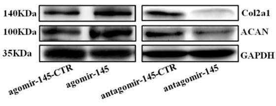
Fig. 3. |miR-145 increased matrix metabolism in rat NP cells.(A, B) Real-time PCR analysis was performed to evaluate the mRNA expression of aggrecan (A) and collagen IIa (B) after a single transfection with agomir-145 or miR-155 inhibitors. (C, D, E) Western blotting (C) and subsequent densitometric analysis (D) demonstrated that aggrecan (C, E) and collagen IIa (D, E) protein were significantly upregulated in rat NP cells after transfection with agomir-145.
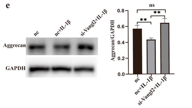
Fig. 4 Silencing of Vangl2 prevents Col-2 and aggrecan degradation in OA. a Western blotting of Col-2 in three groups. The images were quantitatively analyzed and normalized to GAPDH. b The change of Col-2 mRNA was quantitated by RT-qPCR. Representative ICC images of Col-2 (c) and the IOD analysis (d). Scale bar, 50 μm. e The aggrecan expression was measured by Western blotting and quantified for three groups. ns, not statistically significant, *P < 0.05, **P < 0.01; IOD, integrated option density
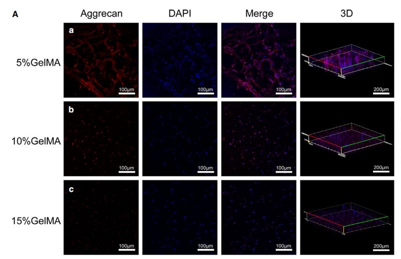
FIGURE 4 Expression of aggrecan of NPCs encapsulated in the GelMA hydrogel. (A) Immunofluorescence staining of NPCs 7 days after encapsulation at different concentrations GelMA hydrogels. (B) Quantitative optical density analysis of aggrecan expression. (*P < .05, **P < .01)
Figure 2. |Protein expression levels of collagen II and aggrecan increase with the added concentration of genistein, while caspase 3 protein levels gradually decrease in each group. (A) Western blot analysis of samples from the experimental groups were tested with the indicated antibodies.
Fig. 3. |LOX improved the ECM expression pattern in TNF-α-treated rat NP cells. A. and B. Relative mRNA expressions of type II collagen and aggrecan in rat NP cells with different treatments were analyzed by RT-qPCR. C. Protein expressions of type II collagen and aggrecan in rat NP cells with different treatments were analyzed by western blot.
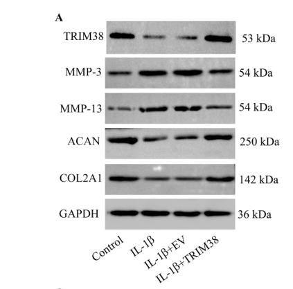
Fig. 4. The effects of TRIM38 depletion on IL-1β-induced degeneration of chondrocytes. (A, B) The effects of TRIM38 overexpression on the levels of MMP-3, MMP- 13, ACAN, and COL2A1 were measured via western blotting (n = 3). (C, D) The effects of TRIM38 knockdown on the levels of MMP-3, MMP-13, ACAN, and COL2A1 were assessed via western blotting (n = 3). **p < 0.01.
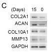
Fig 2 FGF9 expression increased during early differentiation of ATDC5 cells. ATDC5 cells effectively differentiated into chondrogenic cells and stayed at the early stage of chondrogenic differentiation by day 15 as detected by quantitative real-time PCR analysis of the chondrogenic genes Col2, Aggrecan, Col10, Ihh, Mmp13 and FGFR3 Safranin-O staining and Alcian blue staining were carried out on the 15th day and 0th day of induction(B), as well as the protein levels of Col2a1, Acan, Col10a1, Mmp13 detected by Western blot (C). Quantitative real-time PCR (A) and Western blotting analysis (D) showed increased expression of FGF9 during 15 days of ATDC5 differentiation. Immunofluorescence staining (E/a-d) and immunohistochemical staining (E/e) were used to detect the expression and distribution of FGF9 in undifferentiated ATDC5 cells.
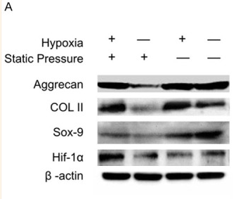
Figure 8 Evaluation of protein expression and cartilage-related genes in the chondrocyte sheets cultured in vitro under static pressure and hypoxic conditions. (A) Protein expression of Aggrecan, COL II, Sox-9, and HIF-1α as determined using western blotting; β-actin was used as a loading control. The expression of Aggrecan (B), COL II (C), Sox-9 (D) and HIF1-α (E) genes was determined using real-time PCR. GAPDH was used as a housekeeping gene. Data are presented as the mean ± standard deviation (n=3),
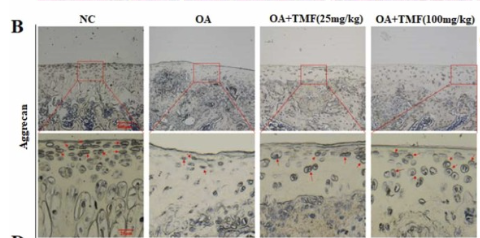
Fig. 1. The effect of TMF on the articular cartilage of OA rats (n = 6) was investigated. (A) The articular cartilage was stained with H/E. IHC assays were used to detect the altered expression of Aggrecan (B–C), ADAMTS5 (D–E), and Caspase3 (F–G) in the cartilage.
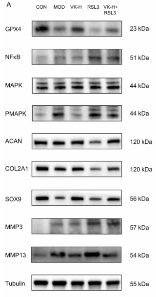
Fig. 8. GPX4 inhibitor RSL3 substantially attenuates VK2 protection of chondrocytes. (A-I) WB results for GPX4, NFκB, pMAPK/MAPK, Aggrecan, CollagenⅡ, SOX9, MMP3, MMP13 and Tubulin; (J) Quantitive analysis of relative Glutathione Peroxidase Activity; (K-L) GSH contents and ratio of GSH/GSSG; (M) Quantitative results of MDA content assay.
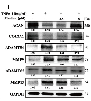
Figure 1 The impact of MA on ECM anabolism and catabolism in TNFα-treated NPCs (A) Chemical architecture of MA. (B) The cytotoxicity of MA to NPCs was evaluated at designated time points by CCK-8 assay at various concentrations. (C–H) The relative mRNA levels of COL2A1, ACAN, ADAMTS5, ADAMTS4, MMP13 and MMP9 were quantified via qPCR. (I) The protein expression levels of ACAN, COL2A1, ADAMTS4, MMP9, ADAMTS5 and MMP13 were assessed in NPCs treated with or without MA in the presence of TNFα. *P
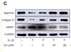
FIGURE 4 Compound 22o alleviates IL-1β-induced chondrocyte inflammatory responses and ECM degradation. Rat chondrocytes were treated with different concentrations of compound 22o in the presence or absence of IL-1β (10 ng/ml) for 24 h. (A) Western blots and (B) quantitative analysis of COX-2. (C) Western blots and (D) quantitative analysis of ACAN, COL2A1 and MMP3. GAPDH was employed as the internal control (n = 3). (E) Quantification of mRNA levels for COX-2, ACAN, COL2A1 and MMP3. Fold changes relative to control group are shown (n = 3). #p < 0.05 vs. control group; *p < 0.05 vs. IL-1β group.
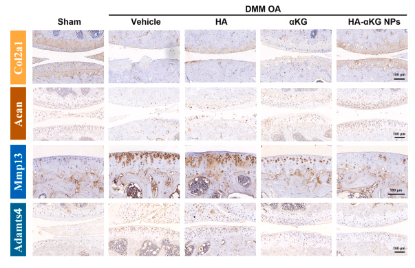
Fig. 9. αKG protects articular cartilage by modulating cartilage matrix homeostasis. Immunohistochemistry staining of Col2a1, Acan, MMP13, and Adamts4 in the knee joint cartilage of advanced OA mice under different treatments.

品牌

产品货号

来源种属

Rabbit

抗体克隆

Polyclonal

来源亚型

IgG

实验方法

WB,IHC,IF,ICC

实验种属

Human,Mouse,Rat,Rabbit,Pig,Dog,Chicken,Bovine,Horse,Sheep

偶联标记

Unconjugated

目的蛋白

Aggrecan

产品规格

50μl,100μl,200μl

产品报价

¥1500/¥2750/¥3600

实验应用

Western blotting

Recommended dilution: 1:1000-3000


Immunofluorescence

Recommended dilution: 1:100-1:500


immunocytochemistry

Recommended dilution: 1:100-1:500


Immunohistochemistry

Recommended dilution: 1:50-1:200


最佳稀释倍数与浓度应由实验研究人员确认

产品说明


产品背景

This proteoglycan is a major component of extracellular matrix of cartilagenous tissues. A major function of this protein is to resist compression in cartilage. It binds avidly to hyaluronic acid via an N-terminal globular region.

Description
Rabbit polyclonal antibody to Aggrecan

Applications 
WB, IF, ICC, IHC.

Immunogen 
Aggrecan Antibody detects endogenous levels of total Aggrecan.

Reactivity 
Human, Mouse, Rat.
可预测:Pig(100%), Bovine(%), Sheep(%), Rabbit(%), Dog(%)

Molecular weight
70,150,250 kDa; 261kD(Calculated).

Host species 
Rabbit

Ig class 
Immunogen-specific rabbit IgG

Purification 
Antigen affinity purification

Full name 
Aggrecan

Synonyms 
ACAN; AGC 1; AGC1; AGCAN; Aggrecan 1 (chondroitin sulfate proteoglycan 1, large aggregating proteoglycan, antigen identified by monoclonal antibody A0122); Aggrecan 1; Aggrecan core protein; Aggrecan proteoglycan; Aggrecan structural proteoglycan of cartilage; Aggrecan1; ATEGQV; Cartilage specific proteoglycan core protein; Chondroitin sulfate proteoglycan 1; Chondroitin sulfate proteoglycan 1 large aggregating proteoglycan antigen identified by monoclonal antibody A0122; Chondroitin sulfate proteoglycan core protein 1; CSPG 1; CSPG1; CSPGCP; JSCATE; Large aggregating proteoglycan; mcspg; mgsk16; MSK 16; MSK16; SEDK;

Storage
Rabbit IgG in phosphate buffered saline , pH 7.4, 150mM NaCl, 0.02% sodium azide and 50% glycerol. Store at -20 °C. Stable for 12 months from date of receipt.

Swissprot 
P16112

产品图片
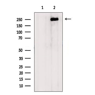
Western blot analysis of extracts from 3T3, using Aggrecan Antibody. The lane on the left was treated with blocking peptide.

ab17521 at 1/100 staining Rat brain tissue by IHC-P. The sample was formaldehyde fixed and a heat mediated antigen retrieval step in citrate buffer was performed. The sample was then blocked and incubated with the primary antibody at 4°C overnight. An HRP conjugated anti-Rabbit antibody was used as the secondary antibody.

ab17521 at 1/100 staining Mouse heart tissue by IHC-P. The sample was formaldehyde fixed and a heat mediated antigen retrieval step in citrate buffer was performed. The sample was then blocked and incubated with the primary antibody at 4°C overnight. An HRP conjugated anti-Rabbit antibody was used as the secondary antibody.

ab17521 at 1/100 staining Mouse brain tissue by IHC-P. The sample was formaldehyde fixed and a heat mediated antigen retrieval step in citrate buffer was performed. The sample was then blocked and incubated with the primary antibody at 4°C overnight. An HRP conjugated anti-Rabbit antibody was used as the secondary antibody.

ab17521 at 1/100 staining Mouse skin tissue by IHC-P. The sample was formaldehyde fixed and a heat mediated antigen retrieval step in citrate buffer was performed. The sample was then blocked and incubated with the primary antibody at 4°C overnight. An HRP conjugated anti-Rabbit antibody was used as the secondary antibody.

ab17521 at 1/100 staining Human normal tissues adjacent to mammary cancer by IHC-P. The sample was formaldehyde fixed and a heat mediated antigen retrieval step in citrate buffer was performed. The sample was then blocked and incubated with the primary antibody at 4°C overnight. An HRP conjugated anti-Rabbit antibody was used as the secondary antibody.

ab17521 at 1/100 staining Human mammary cancer by IHC-P. The sample was formaldehyde fixed and a heat mediated antigen retrieval step in citrate buffer was performed. The sample was then blocked and incubated with the primary antibody at 4°C overnight. An HRP conjugated anti-Rabbit antibody was used as the secondary antibody.

Fig. 3. |miR-145 increased matrix metabolism in rat NP cells.(A, B) Real-time PCR analysis was performed to evaluate the mRNA expression of aggrecan (A) and collagen IIa (B) after a single transfection with agomir-145 or miR-155 inhibitors. (C, D, E) Western blotting (C) and subsequent densitometric analysis (D) demonstrated that aggrecan (C, E) and collagen IIa (D, E) protein were significantly upregulated in rat NP cells after transfection with agomir-145.

Fig. 4 Silencing of Vangl2 prevents Col-2 and aggrecan degradation in OA. a Western blotting of Col-2 in three groups. The images were quantitatively analyzed and normalized to GAPDH. b The change of Col-2 mRNA was quantitated by RT-qPCR. Representative ICC images of Col-2 (c) and the IOD analysis (d). Scale bar, 50 μm. e The aggrecan expression was measured by Western blotting and quantified for three groups. ns, not statistically significant, *P < 0.05, **P < 0.01; IOD, integrated option density

FIGURE 4 Expression of aggrecan of NPCs encapsulated in the GelMA hydrogel. (A) Immunofluorescence staining of NPCs 7 days after encapsulation at different concentrations GelMA hydrogels. (B) Quantitative optical density analysis of aggrecan expression. (*P < .05, **P < .01)

Figure 2. |Protein expression levels of collagen II and aggrecan increase with the added concentration of genistein, while caspase 3 protein levels gradually decrease in each group. (A) Western blot analysis of samples from the experimental groups were tested with the indicated antibodies.

Fig. 3. |LOX improved the ECM expression pattern in TNF-α-treated rat NP cells. A. and B. Relative mRNA expressions of type II collagen and aggrecan in rat NP cells with different treatments were analyzed by RT-qPCR. C. Protein expressions of type II collagen and aggrecan in rat NP cells with different treatments were analyzed by western blot.

Fig. 4. The effects of TRIM38 depletion on IL-1β-induced degeneration of chondrocytes. (A, B) The effects of TRIM38 overexpression on the levels of MMP-3, MMP- 13, ACAN, and COL2A1 were measured via western blotting (n = 3). (C, D) The effects of TRIM38 knockdown on the levels of MMP-3, MMP-13, ACAN, and COL2A1 were assessed via western blotting (n = 3). **p < 0.01.

Fig 2 FGF9 expression increased during early differentiation of ATDC5 cells. ATDC5 cells effectively differentiated into chondrogenic cells and stayed at the early stage of chondrogenic differentiation by day 15 as detected by quantitative real-time PCR analysis of the chondrogenic genes Col2, Aggrecan, Col10, Ihh, Mmp13 and FGFR3 Safranin-O staining and Alcian blue staining were carried out on the 15th day and 0th day of induction(B), as well as the protein levels of Col2a1, Acan, Col10a1, Mmp13 detected by Western blot (C). Quantitative real-time PCR (A) and Western blotting analysis (D) showed increased expression of FGF9 during 15 days of ATDC5 differentiation. Immunofluorescence staining (E/a-d) and immunohistochemical staining (E/e) were used to detect the expression and distribution of FGF9 in undifferentiated ATDC5 cells.

Figure 8 Evaluation of protein expression and cartilage-related genes in the chondrocyte sheets cultured in vitro under static pressure and hypoxic conditions. (A) Protein expression of Aggrecan, COL II, Sox-9, and HIF-1α as determined using western blotting; β-actin was used as a loading control. The expression of Aggrecan (B), COL II (C), Sox-9 (D) and HIF1-α (E) genes was determined using real-time PCR. GAPDH was used as a housekeeping gene. Data are presented as the mean ± standard deviation (n=3),

Fig. 1. The effect of TMF on the articular cartilage of OA rats (n = 6) was investigated. (A) The articular cartilage was stained with H/E. IHC assays were used to detect the altered expression of Aggrecan (B–C), ADAMTS5 (D–E), and Caspase3 (F–G) in the cartilage.

Fig. 8. GPX4 inhibitor RSL3 substantially attenuates VK2 protection of chondrocytes. (A-I) WB results for GPX4, NFκB, pMAPK/MAPK, Aggrecan, CollagenⅡ, SOX9, MMP3, MMP13 and Tubulin; (J) Quantitive analysis of relative Glutathione Peroxidase Activity; (K-L) GSH contents and ratio of GSH/GSSG; (M) Quantitative results of MDA content assay.

Figure 1 The impact of MA on ECM anabolism and catabolism in TNFα-treated NPCs (A) Chemical architecture of MA. (B) The cytotoxicity of MA to NPCs was evaluated at designated time points by CCK-8 assay at various concentrations. (C–H) The relative mRNA levels of COL2A1, ACAN, ADAMTS5, ADAMTS4, MMP13 and MMP9 were quantified via qPCR. (I) The protein expression levels of ACAN, COL2A1, ADAMTS4, MMP9, ADAMTS5 and MMP13 were assessed in NPCs treated with or without MA in the presence of TNFα. *P

FIGURE 4 Compound 22o alleviates IL-1β-induced chondrocyte inflammatory responses and ECM degradation. Rat chondrocytes were treated with different concentrations of compound 22o in the presence or absence of IL-1β (10 ng/ml) for 24 h. (A) Western blots and (B) quantitative analysis of COX-2. (C) Western blots and (D) quantitative analysis of ACAN, COL2A1 and MMP3. GAPDH was employed as the internal control (n = 3). (E) Quantification of mRNA levels for COX-2, ACAN, COL2A1 and MMP3. Fold changes relative to control group are shown (n = 3). #p < 0.05 vs. control group; *p < 0.05 vs. IL-1β group.

Fig. 9. αKG protects articular cartilage by modulating cartilage matrix homeostasis. Immunohistochemistry staining of Col2a1, Acan, MMP13, and Adamts4 in the knee joint cartilage of advanced OA mice under different treatments.

联系经销商
北京晶莱华科生物技术有限公司
北京总部电话:4008-766-085
邮箱:beijing@genlay.cn
地址:北京经济技术开发区科创十三街12号院德为科技园1号楼5层
上海分公司电话:4008-766-085
邮箱:shanghai@genlay.cn
地址:上海市青浦区普仓路648弄71号
长沙分公司电话:4008-766-085
邮箱:changsha@genlay.cn
地址:长沙市岳麓区麓谷大道627号新长海中心A1栋1203室
http://www.genlay.cn
http://www.genecro.cn
genetex@neobioscience.com