ab11101 AGER/RAGE Antibody

Western blot analysis of extracts from Rat lung, using AGER(RAGE) Antibody. The lane on the left was treated with blocking peptide.
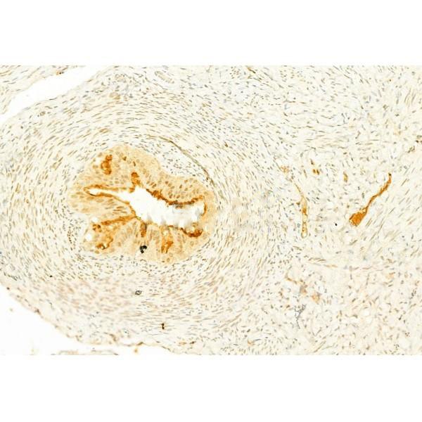
ab11101 at 1/100 staining Rat ovary tissue by IHC-P. The sample was formaldehyde fixed and a heat mediated antigen retrieval step in citrate buffer was performed. The sample was then blocked and incubated with the primary antibody at 4°C overnight. An HRP conjugated anti-Rabbit antibody was used as the secondary antibody.
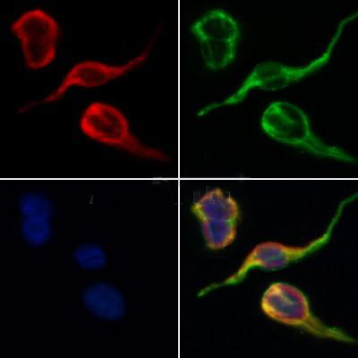
ab11101 staining HepG2 by IF/ICC. The samples were fixed with PFA and permeabilized in 0.1% Triton X-100,then blocked in 10% serum for 45 minutes at 25°C. Samples were then incubated with primary Ab(ab11101 1:200) and mouse anti-beta tubulin Ab(T0023 1:200) for 1 hour at 37°C. An AlexaFluor594 conjugated goat anti-rabbit IgG(H+L) Ab(Red) and an AlexaFluor488 conjugated goat anti-mouse IgG(H+L) Ab(Green) were used as the secondary antibody. The nuclear counter stain is DAPI(blue).
Figure 1 |HMGB1 knockdown decreased the viability of A498 and ACHN cells.Notes: (A) The RAGE and HMGB1 proteins in RCC cell lines were detected by Western blot.
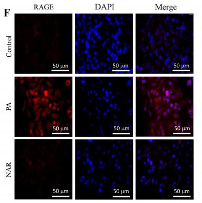
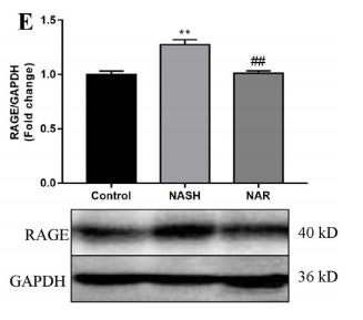
Fig. 4. |Naringin ameliorates NASH through modulation of the AGE/RAGE mechanism. The level of AGE was determined in HepG2 cells and plasma by (A) ELISA and(B) fluorimetry. Receptor for AGE (RAGE) was analyzed in HepG2 cells and liver tissue by (C) qPCR (n = 6) and (D and E) immunoblotting (n = 3). Expressions were normalized by GAPDH. RAGE expression was further validated by immunofluorescence in (F) cells, (G) its quantification (n = 5), and (H) tissue and (I) its quantification (n = 5) by using Alexa Fluor 594 (red), DAPI (blue). Magnification 40×. Data are shown as Mean ± S.E.M (n = 8), *Control vs NASH; #NASH vs NAR. **P < 0.01, ***P < 0.001; #P < 0.05, ##P < 0.01, ###P < 0.001.
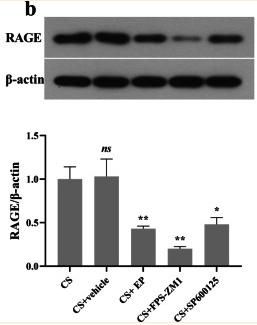
Fig. 2 EP, FPS-ZM1, and SP600125 affect the expression of HMGB1, RAGE, and JNK in lung tissue. Relative levels of HMGB1, RAGE, and JNK were analyzed using Western blot. The HMGB1, RAGE, and p-JNK were lower in the later three pretreatment groups. Results were expressed as means ± SDs. The number of rats were 13, 12, 14, 13, and 14 in CS, CS + vehicle, CS + EP, CS + FPS-ZM1, and CS + SP600125 groups, respectively. Statistical significance: ns P ≥ 0.05, *P < 0.05 and **P < 0.01, versus CS group

品牌

产品货号

来源种属

Rabbit

抗体克隆

Polyclonal

来源亚型

IgG

实验方法

WB,IHC,IF,ICC

实验种属

Human,Mouse,Rat,Rabbit,Pig,Dog,Chicken,Bovine,Horse,Sheep

偶联标记

Unconjugated

目的蛋白

AGER/RAGE

产品规格

50μl,100μl,200μl

产品报价

¥1500/¥2750/¥3600

实验应用

Western blotting

Recommended dilution: 1:500-1:2000


Immunofluorescence

Recommended dilution: 1:100-1:500


immunocytochemistry

Recommended dilution: 1:100-1:500


Immunohistochemistry

Recommended dilution: 1:50-1:200


最佳稀释倍数与浓度应由实验研究人员确认

产品说明


产品背景

Mediates interactions of advanced glycosylation end products (AGE). These are nonenzymatically glycosylated proteins which accumulate in vascular tissue in aging and at an accelerated rate in diabetes. Acts as a mediator of both acute and chronic vascular inflammation in conditions such as atherosclerosis and in particular as a complication of diabetes. AGE/RAGE signaling plays an important role in regulating the production/expression of TNF-alpha, oxidative stress, and endothelial dysfunction in type 2 diabetes. Interaction with S100A12 on endothelium, mononuclear phagocytes, and lymphocytes triggers cellular activation, with generation of key proinflammatory mediators. Interaction with S100B after myocardial infarction may play a role in myocyte apoptosis by activating ERK1/2 and p53/TP53 signaling (By similarity). Receptor for amyloid beta peptide. Contributes to the translocation of amyloid-beta peptide (ABPP) across the cell membrane from the extracellular to the intracellular space in cortical neurons. ABPP-initiated RAGE signaling, especially stimulation of p38 mitogen-activated protein kinase (MAPK), has the capacity to drive a transport system delivering ABPP as a complex with RAGE to the intraneuronal space. Can also bind oligonucleotides.

Description
Rabbit polyclonal antibody to AGER/RAGE

Applications 
WB, IF, ICC, IHC

Immunogen 
AGER/RAGE Antibody detects endogenous levels of total AGER/RAGE.

Reactivity 
Human, Mouse, Rat.
可预测:Pig(92%), Bovine(%), Horse(%), Sheep(%), Rabbit(%), Dog(%)

Molecular weight
43~60kDa; 43kD(Calculated).

Host species 
Rabbit

Ig class 
Immunogen-specific rabbit IgG

Purification 
Antigen affinity purification

Full name 
AGER/RAGE

Synonyms 
Advanced glycosylation end product-specific receptor; Ager; MGC2235; RAGE_HUMAN; Receptor for advanced glycosylation end products;

Storage
Rabbit IgG in phosphate buffered saline , pH 7.4, 150mM NaCl, 0.02% sodium azide and 50% glycerol. Store at -20 °C. Stable for 12 months from date of receipt.

Swissprot 
Q15109

产品图片
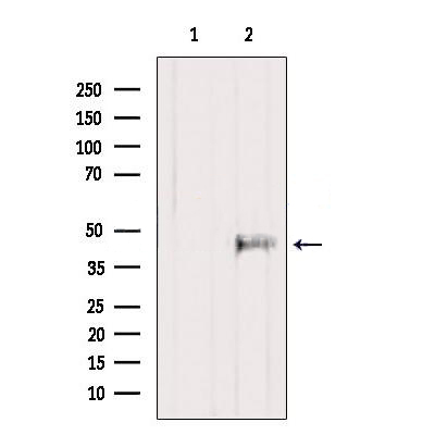
Western blot analysis of extracts from Rat lung, using AGER(RAGE) Antibody. The lane on the left was treated with blocking peptide.

ab11101 at 1/100 staining Rat ovary tissue by IHC-P. The sample was formaldehyde fixed and a heat mediated antigen retrieval step in citrate buffer was performed. The sample was then blocked and incubated with the primary antibody at 4°C overnight. An HRP conjugated anti-Rabbit antibody was used as the secondary antibody.

ab11101 staining HepG2 by IF/ICC. The samples were fixed with PFA and permeabilized in 0.1% Triton X-100,then blocked in 10% serum for 45 minutes at 25°C. Samples were then incubated with primary Ab(ab11101 1:200) and mouse anti-beta tubulin Ab(T0023 1:200) for 1 hour at 37°C. An AlexaFluor594 conjugated goat anti-rabbit IgG(H+L) Ab(Red) and an AlexaFluor488 conjugated goat anti-mouse IgG(H+L) Ab(Green) were used as the secondary antibody. The nuclear counter stain is DAPI(blue).

Figure 1 |HMGB1 knockdown decreased the viability of A498 and ACHN cells.Notes: (A) The RAGE and HMGB1 proteins in RCC cell lines were detected by Western blot.

Fig. 4. |Naringin ameliorates NASH through modulation of the AGE/RAGE mechanism. The level of AGE was determined in HepG2 cells and plasma by (A) ELISA and(B) fluorimetry. Receptor for AGE (RAGE) was analyzed in HepG2 cells and liver tissue by (C) qPCR (n = 6) and (D and E) immunoblotting (n = 3). Expressions were normalized by GAPDH. RAGE expression was further validated by immunofluorescence in (F) cells, (G) its quantification (n = 5), and (H) tissue and (I) its quantification (n = 5) by using Alexa Fluor 594 (red), DAPI (blue). Magnification 40×. Data are shown as Mean ± S.E.M (n = 8), *Control vs NASH; #NASH vs NAR. **P < 0.01, ***P < 0.001; #P < 0.05, ##P < 0.01, ###P < 0.001.

Fig. 4. |Naringin ameliorates NASH through modulation of the AGE/RAGE mechanism. The level of AGE was determined in HepG2 cells and plasma by (A) ELISA and(B) fluorimetry. Receptor for AGE (RAGE) was analyzed in HepG2 cells and liver tissue by (C) qPCR (n = 6) and (D and E) immunoblotting (n = 3). Expressions were normalized by GAPDH. RAGE expression was further validated by immunofluorescence in (F) cells, (G) its quantification (n = 5), and (H) tissue and (I) its quantification (n = 5) by using Alexa Fluor 594 (red), DAPI (blue). Magnification 40×. Data are shown as Mean ± S.E.M (n = 8), *Control vs NASH; #NASH vs NAR. **P < 0.01, ***P < 0.001; #P < 0.05, ##P < 0.01, ###P < 0.001.

Fig. 2 EP, FPS-ZM1, and SP600125 affect the expression of HMGB1, RAGE, and JNK in lung tissue. Relative levels of HMGB1, RAGE, and JNK were analyzed using Western blot. The HMGB1, RAGE, and p-JNK were lower in the later three pretreatment groups. Results were expressed as means ± SDs. The number of rats were 13, 12, 14, 13, and 14 in CS, CS + vehicle, CS + EP, CS + FPS-ZM1, and CS + SP600125 groups, respectively. Statistical significance: ns P ≥ 0.05, *P < 0.05 and **P < 0.01, versus CS group

联系经销商
北京晶莱华科生物技术有限公司
北京总部电话:4008-766-085
邮箱:beijing@genlay.cn
地址:北京经济技术开发区科创十三街12号院德为科技园1号楼5层
上海分公司电话:4008-766-085
邮箱:shanghai@genlay.cn
地址:上海市青浦区普仓路648弄71号
长沙分公司电话:4008-766-085
邮箱:changsha@genlay.cn
地址:长沙市岳麓区麓谷大道627号新长海中心A1栋1203室
http://www.genlay.cn
http://www.genecro.cn
genetex@neobioscience.com